Vansh Diagnostics Akola — Sonography, Digital X-Ray & Fetal Medicine Centre
89995 54391 Book a scan
Akola · Est. 2022
वंश डायग्नोस्टिक

Gentle imaging,
clear answers.

Sonography, digital X-ray, 2D echo and fetal medicine under one calm roof — with same-day reports and gentle, unhurried care.
Book a scan See our services
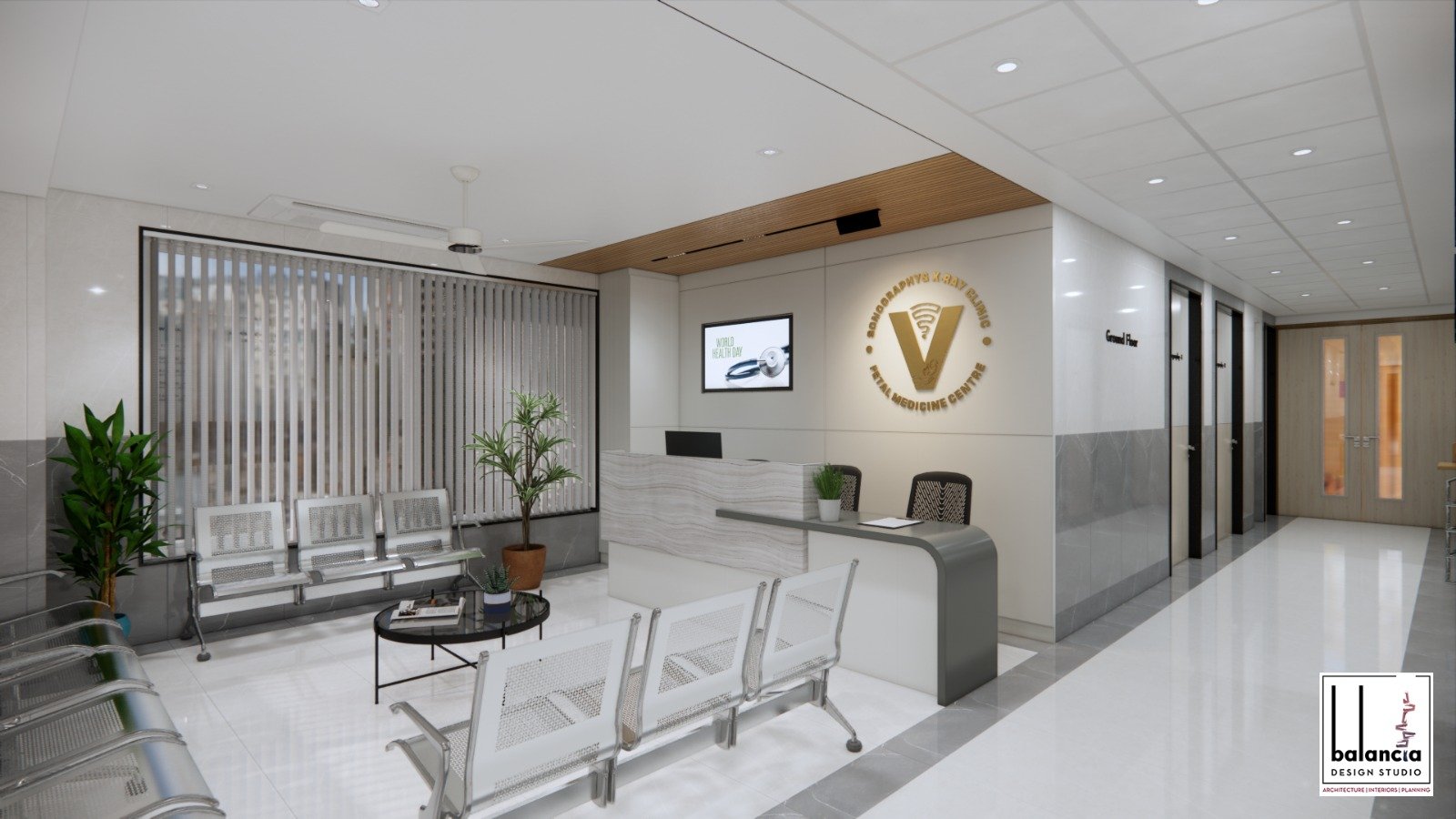
Vansh Diagnostics reception
PCPNDT compliant
Registered & regulated centre
Dr. Sonam Heda (Laddha), MD Radiodiagnosis Fellow in Fetal Medicine MMC 2011/03/0536 PCPNDT Reg. MH-AKL/DI/PNDT/163
{{ svc.icon }}

{{ svc.title }}

{{ svc.description }}

{{ pt }}
Rate list

Transparent charges for every test

See the full X-ray, sonography and colour Doppler rate list — and get a secure payment link on WhatsApp for any test.
View rates & tests
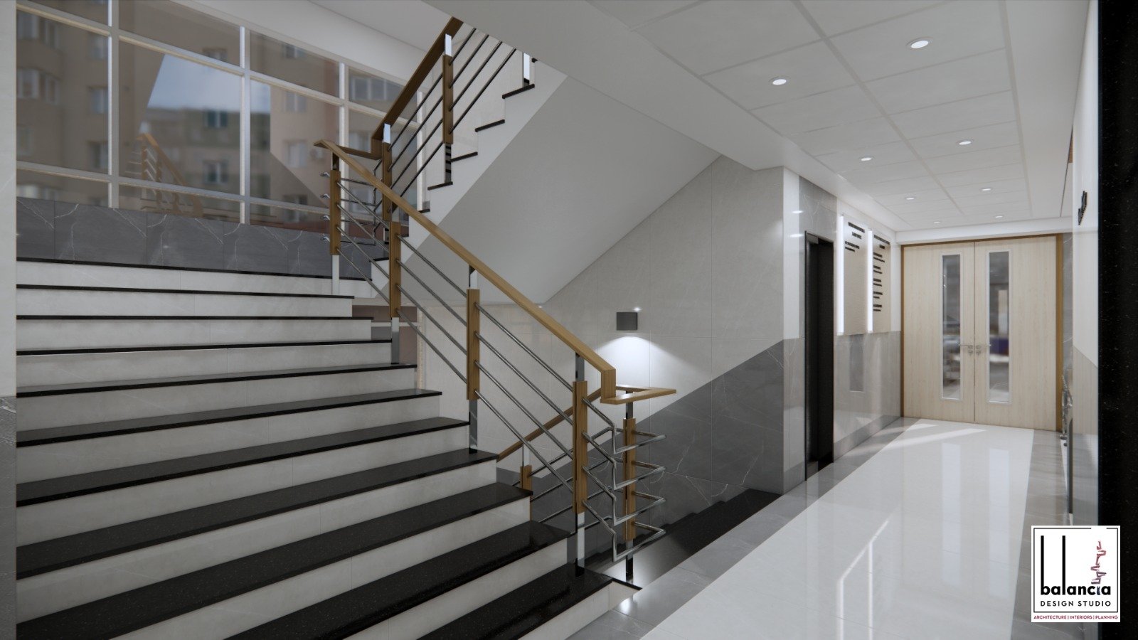
Vansh Diagnostics interior

Vansh Diagnostics was built around a simple idea: imaging should feel calm and understandable. Our purpose-built centre on Main Road brings sonography, digital X-ray, fetal medicine and genetic counselling together — so families get answers quickly, and kindly.
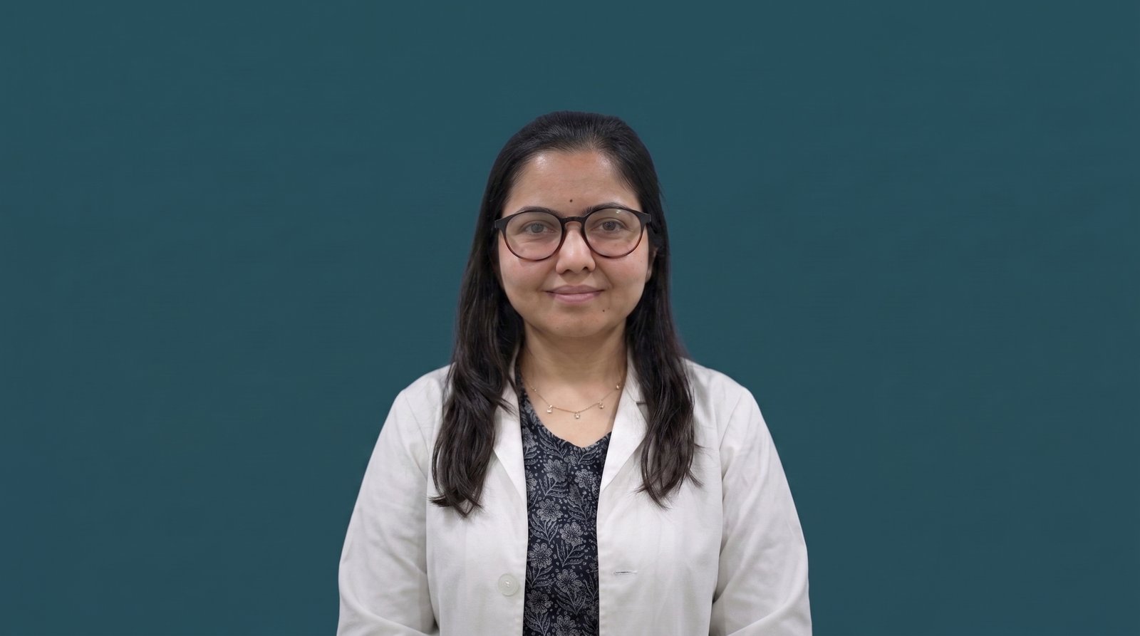
Dr. Sonam Heda (Laddha)
Radiology Head & Fetal Medicine
Dr. Sonam Heda (Laddha)
MBBS, MD Radiodiagnosis · Fellowship in Fetal Medicine · MMC 2011/03/0536

Radiologist with a fellowship in fetomaternal medicine, reporting CT, MRI, ultrasonography and advanced fetal imaging across hospitals in Nagpur, Mumbai, Ludhiana and Kochi — consultant radiologist and fetal medicine expert in Akola since 2021.
Education
MBBS · JNMC, Sawangi (M), Wardha (DMIMS) — 2011
MD Radiodiagnosis · N.K.P. Salve Medical College, Nagpur (MUHS) — 2014
Fellowship in Fetomaternal Medicine · CIMAR Hospital, Ernakulam — 2020
Experience
2014–15 · Senior Resident, Lata Mangeshkar Hospital, Nagpur
2015–16 · Asst. Professor, Grant Medical College & JJ Hospital, Mumbai
2016–17 · Consultant Radiologist, Ludhiana (Ace, Heart Beat Diagnostics)
2018–19 · Consultant Radiologist, Spandan Hospital, Dharmavaram
2019–20 · Consultant Radiologist, Imaging Point
2020–21 · Fetal Medicine, CIMAR, Kochi
Since 2021 · Consultant Radiologist & Fetal Medicine Expert, Akola
Conferences & achievements
65th Annual Conference, Indian Radiological & Imaging Association — Hyderabad, 2012
66th Annual Conference, IRIA — paper/poster presentation — Indore, 2013
CME & Workshop on Obstetrics & Gynaecology, IRIA Vidarbha — Nagpur, 2013
37th State Conference, IRIA — poster on Hemoptysis cases — Nagpur, 2014
MUSKCON — Annual Conference, Karnataka Ultrasound Society — Bengaluru, 2020
Dr. Naresh Tayde
Dr. Naresh Tayde · Visiting geneticist
Geneticist & Paediatrician, Amravati · 2D Echo (paediatric & adult)
Mar 2003 – Apr 2006 · B. J. Medical College & Sassoon Hospital, Dept. of Pediatrics, Pune — JR & SR
Mar 2007 – Feb 2010 · Deenanath Mangeshkar Hospital, Pediatrics, Pune — Medical Doctor
Genetic consultations: first Sunday of every month
Same-day reporting
Most reports are ready before you leave the centre.
Advanced equipment
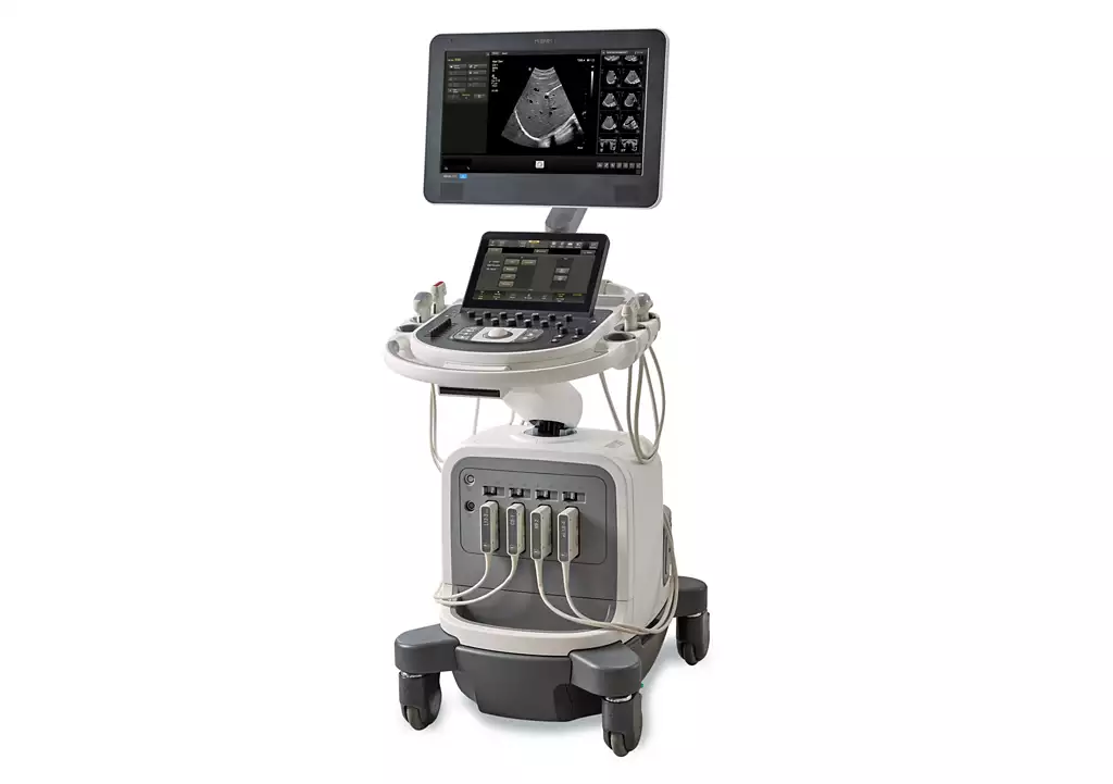
Philips Affinity 70 & ClearVue 360 ultrasound, Adonis 300mA digital X-ray, 2D echo.
Modern, comfortable spaces
Purpose-built rooms designed for privacy and ease.
Philips Affinity 70 ultrasound machine

Philips Affinity 70

Flagship 3D/4D ultrasound — premium image quality for fetal, cardiac and adult imaging
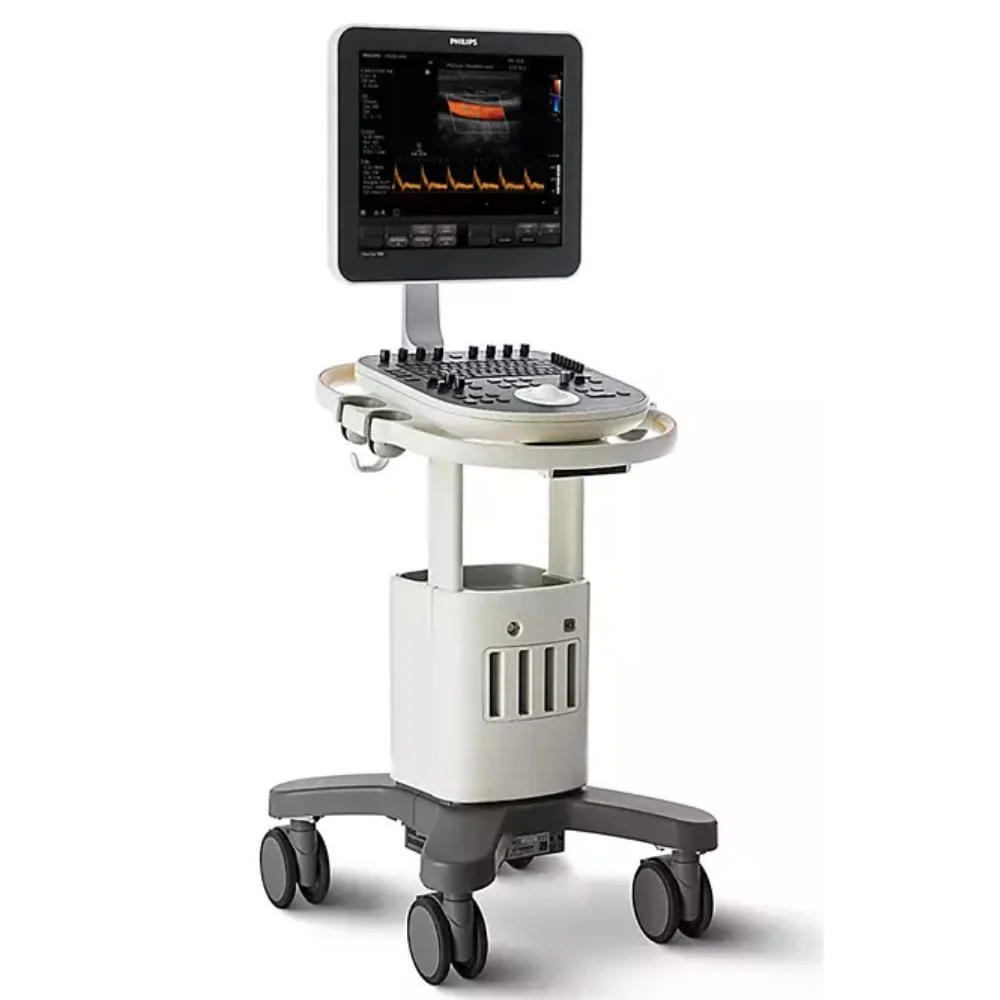
Philips ClearVeu 350 ultrasound machine

Philips ClearVeu 350

Versatile ultrasound platform with colour, pulsed and tissue Doppler
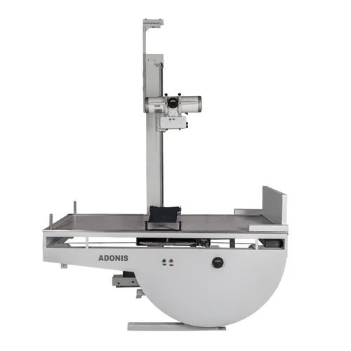
Adonis 300mA digital X-ray machine

Adonis 300mA X-ray

Fixed digital X-ray unit with low-dose imaging and fast reporting
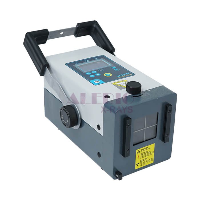
Alerio portable X-ray machine

Alerio Portable X-ray

Home-based portable X-ray service (30Amp) for bedside imaging
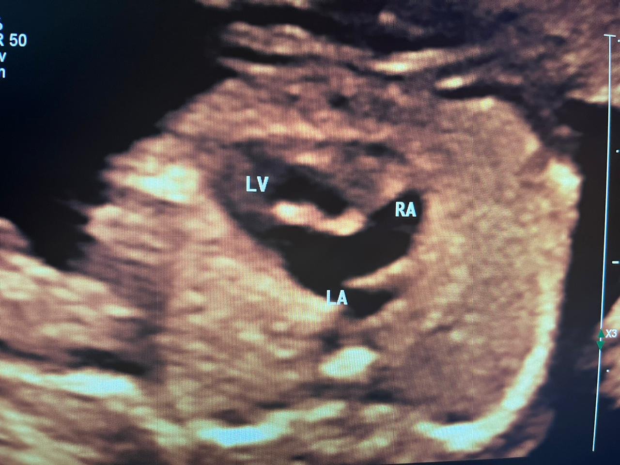
Fetal echocardiography showing DILV with unbalanced AVSD — labelled LV, RA, LA
Fetal echo

DILV / unbalanced AVSD — monoventricle

Double-inlet left ventricle with unbalanced AV septal defect diagnosed on fetal echocardiography — a rare single-ventricle heart, identified before birth.

Serial imaging

Diaphragmatic hernia — progressive presentation

Initial scan: unilateral pleural effusion. Repeat scan 10 days later: progression to diaphragmatic hernia. Key lesson: check SMA position with effusion to identify early hernia.

Ultrasound at 22 weeks showing diaphragmatic eventration / hernia
Fetal medicine

Eventration / hernia at 22 weeks

Diaphragmatic eventration / hernia diagnosed on the 22-week anomaly scan — early detection that lets the family plan delivery and neonatal care in advance.

Congenital heart

Tricuspid atresia with muscular VSD

Critical congenital heart defect diagnosed on fetal echocardiography. Absence of the tricuspid valve with muscular ventricular septal defect requiring immediate postnatal intervention.

Cardiac

Cardiac rhabdomyoma — 3rd trimester

Benign cardiac tumour diagnosed on fetal echocardiography in the third trimester — follow-up imaging confirmed resolution postnatally.

Syndromic

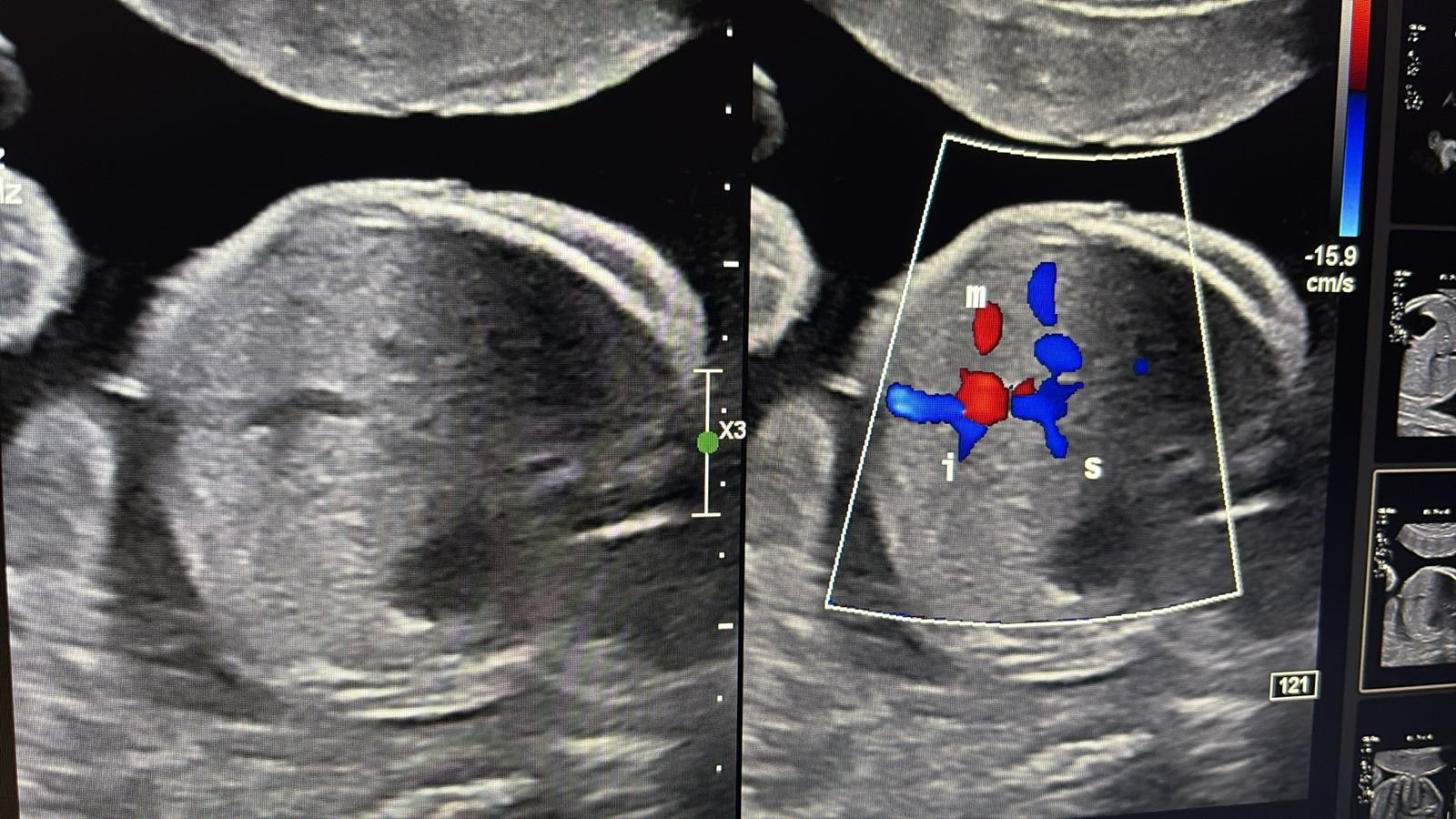
Klippel-Trenaunay syndrome — cystic neck mass

Solid-cystic neck mass identified on the 21-week anomaly scan. Klippel-Trenaunay syndrome involving vascular malformation and soft tissue overgrowth requiring coordinated postnatal management.

Genetics

Trisomy 21 — first trimester NT pick

Early detection of Down syndrome on the 12-week nuchal translucency scan. Timely identification enabling informed family decision-making and antenatal care planning.

Grateful to mentor Dr. Bijoy Balakrishnan for guidance on this case
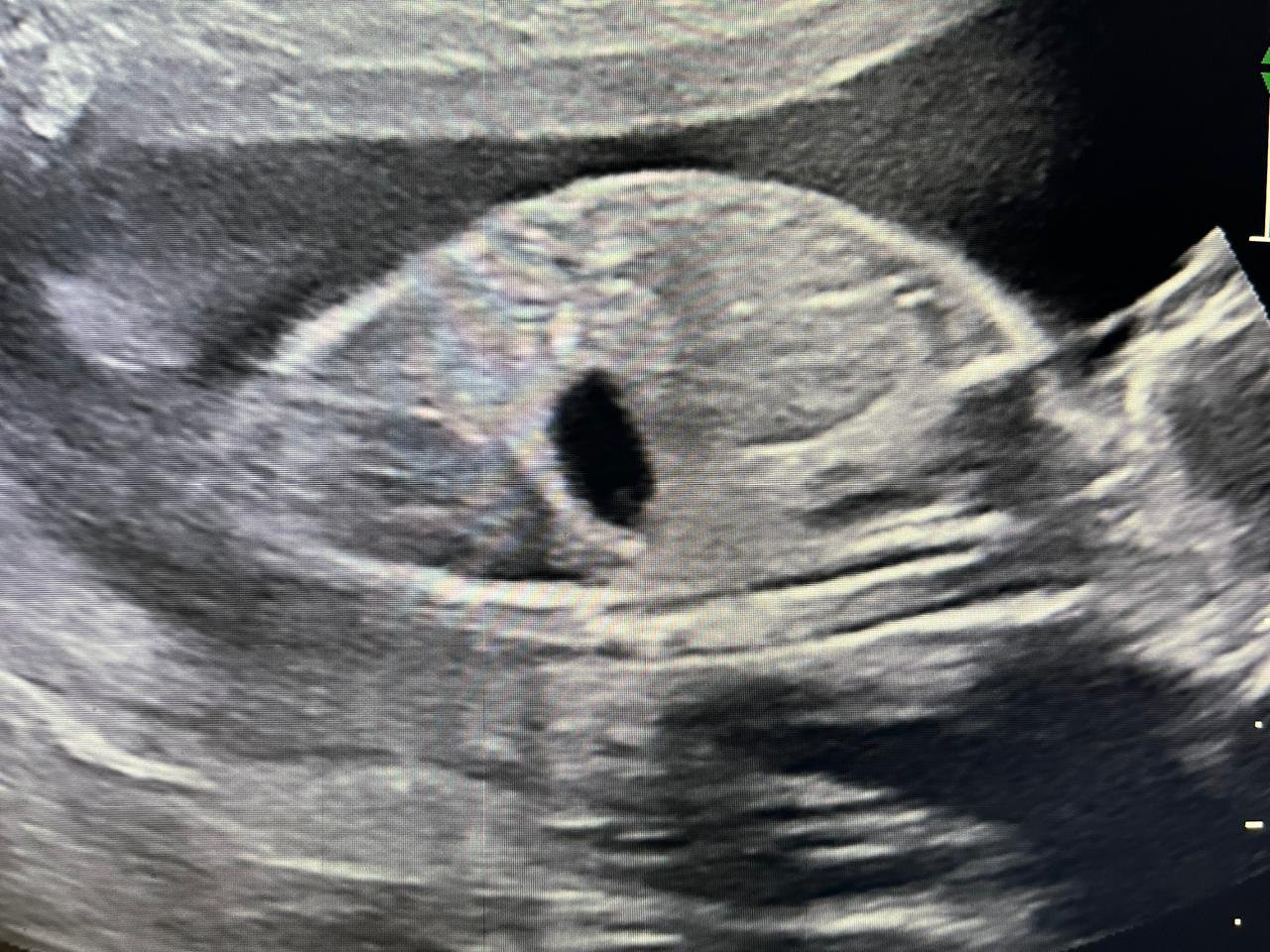
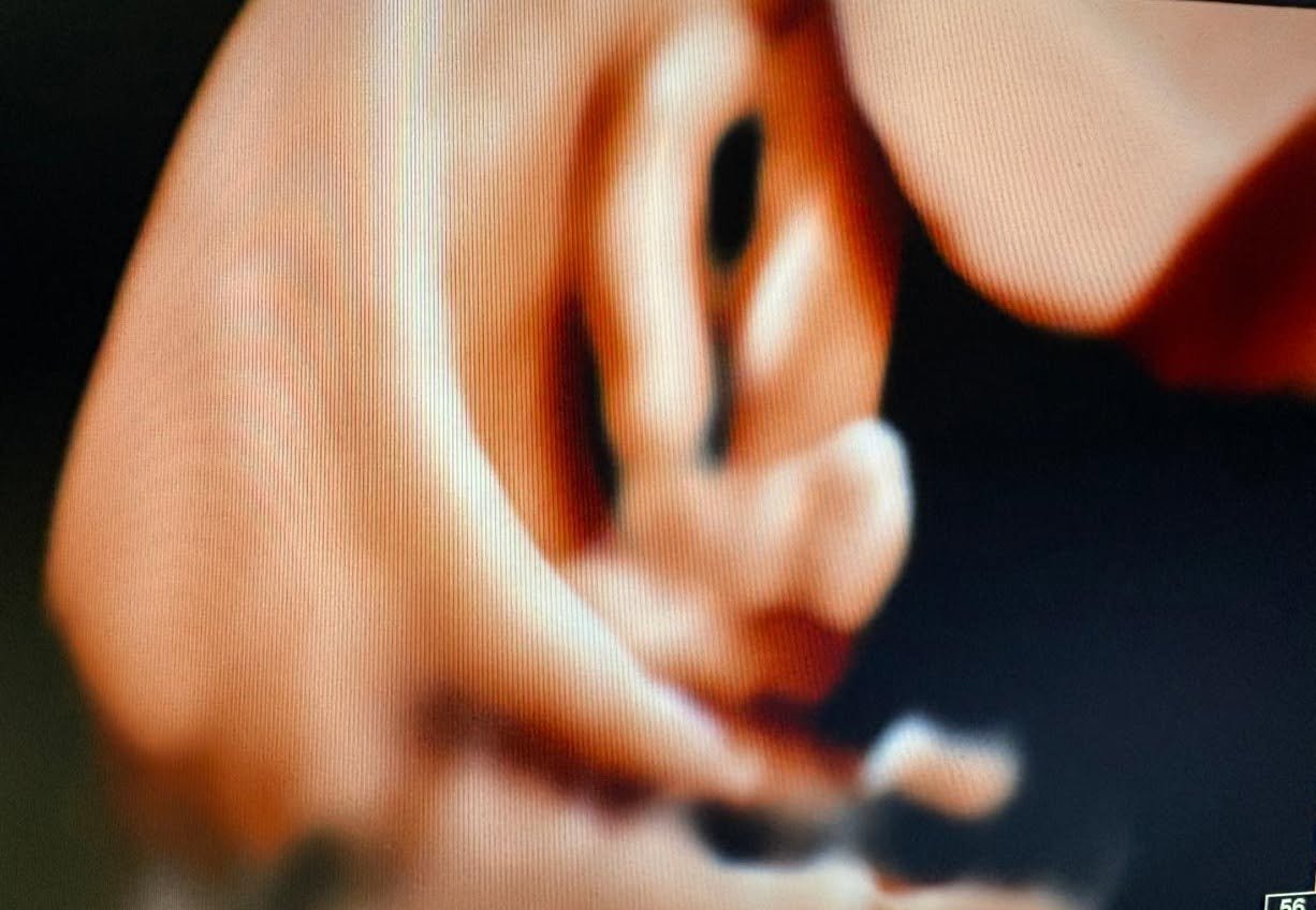
Antenatal ultrasound showing congenital club foot
Fetal medicine

Club foot picked up at 12 weeks on NT scan

Congenital talipes identified as early as the 12-week nuchal translucency scan — letting the family plan corrective treatment right from birth.

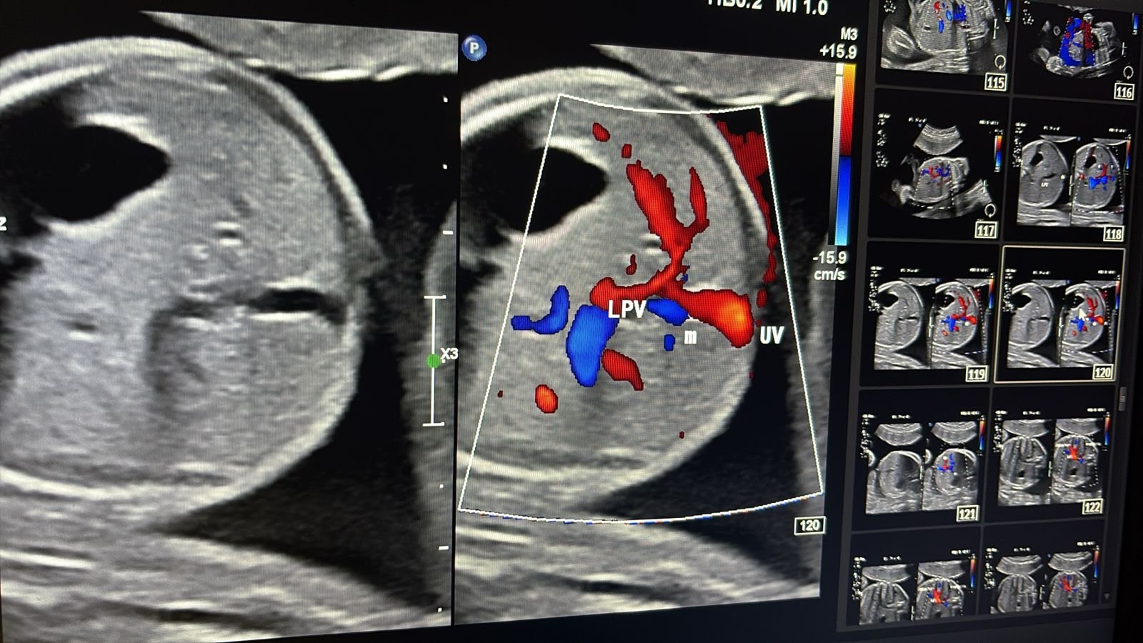
Colour Doppler showing normal fetal portal venous system — LPV and UV
Normal anatomy

Normal fetal portal venous system

Colour Doppler mapping of the normal portal venous anatomy — left and right portal veins, umbilical vein and their branching pattern demonstrated on the anomaly scan.

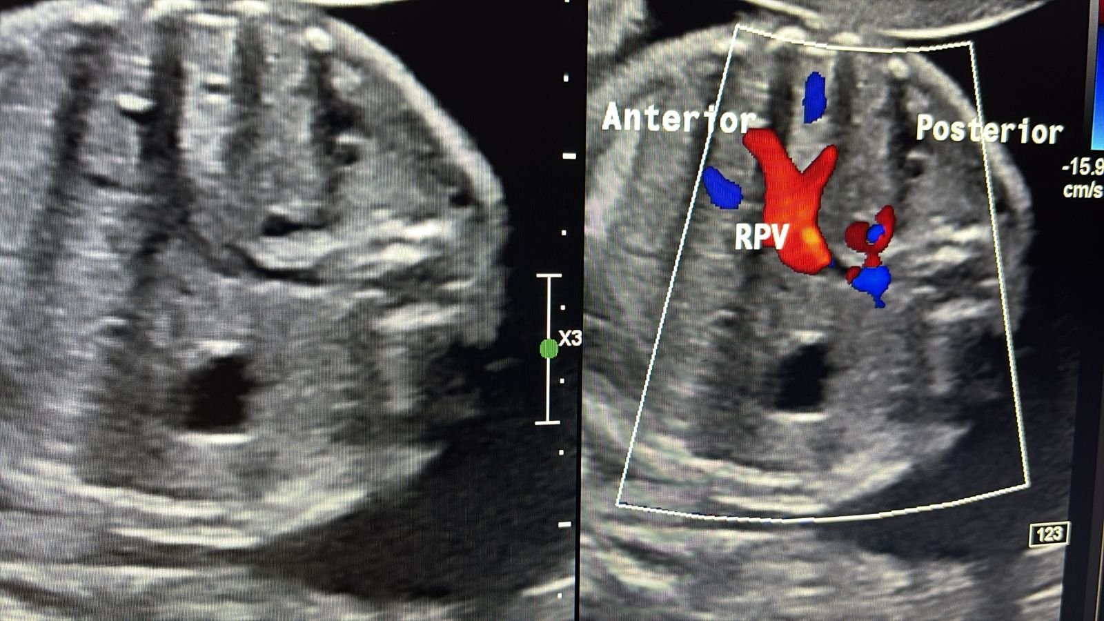
Right portal vein on colour Doppler Portal venous branches on colour Doppler
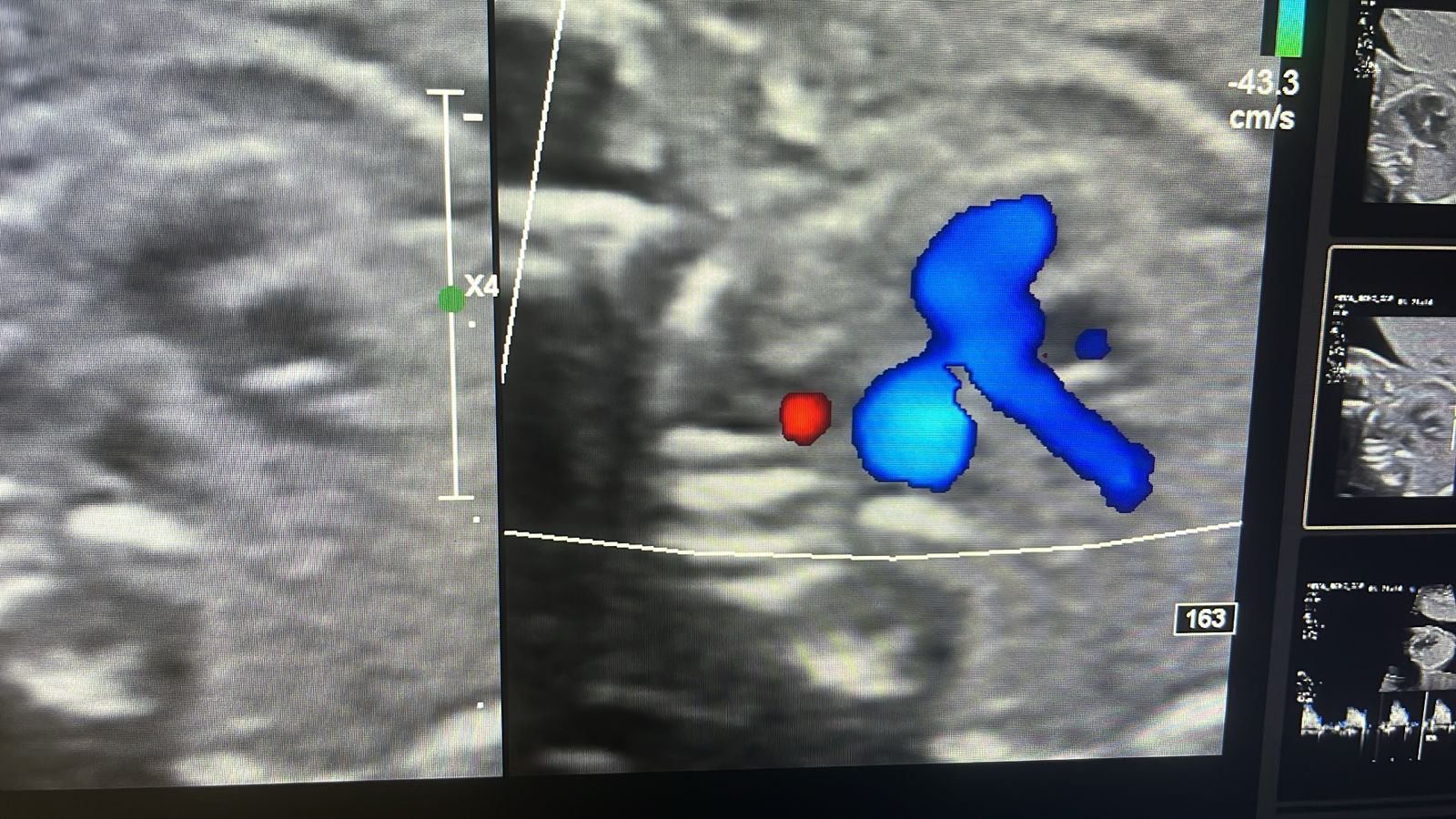
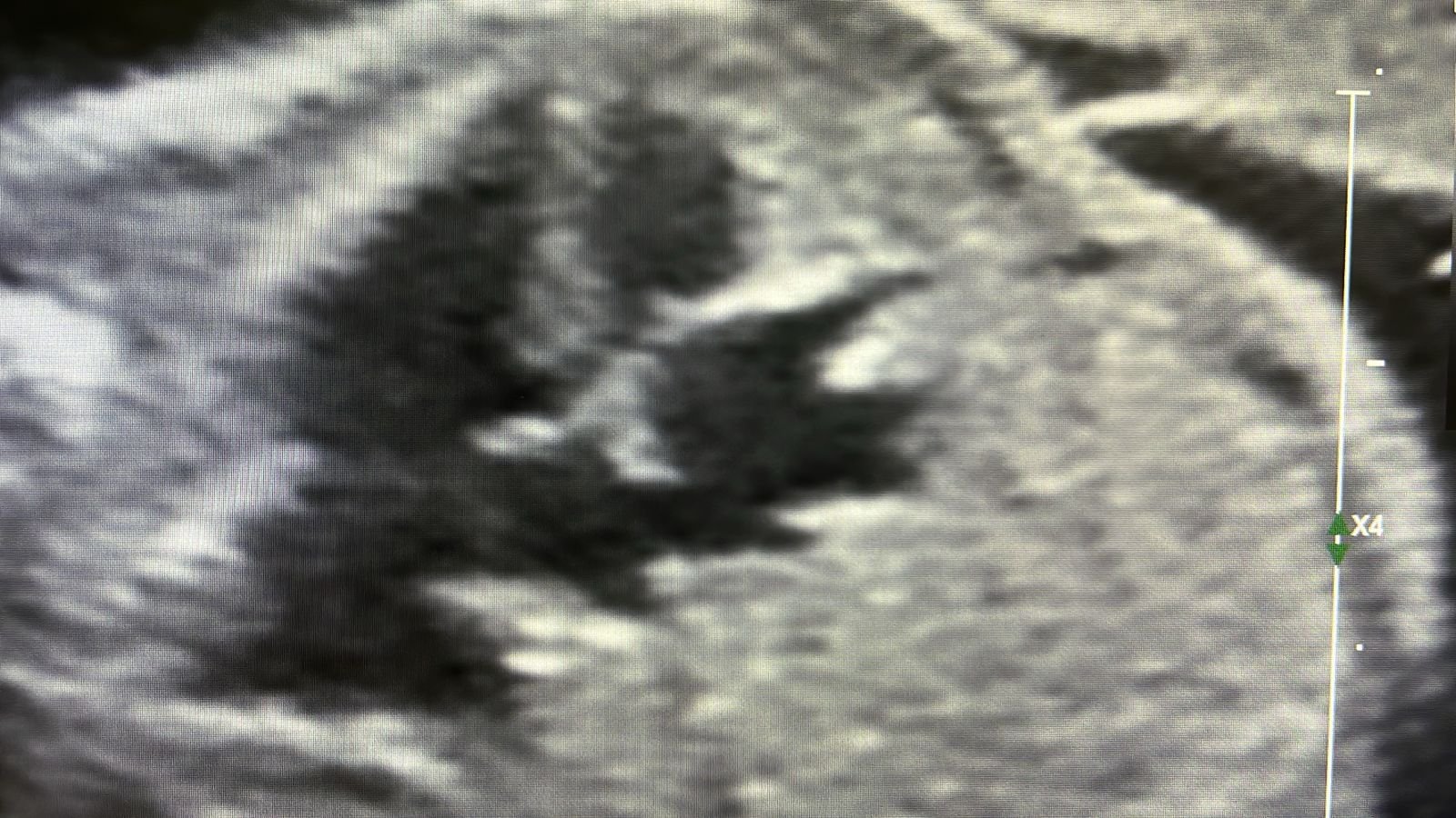
Horseshoe kidney on colour Doppler at 21 weeks Horseshoe kidney sagittal view at 21 weeks
Renal

Horseshoe kidney — fetal anomaly scan

Pelvic pancake / horseshoe kidney variant diagnosed on the 21-week anatomy scan, with associated pulmonary hypoplasia. Early detection enabling postnatal follow-up and appropriate neonatal management planning.

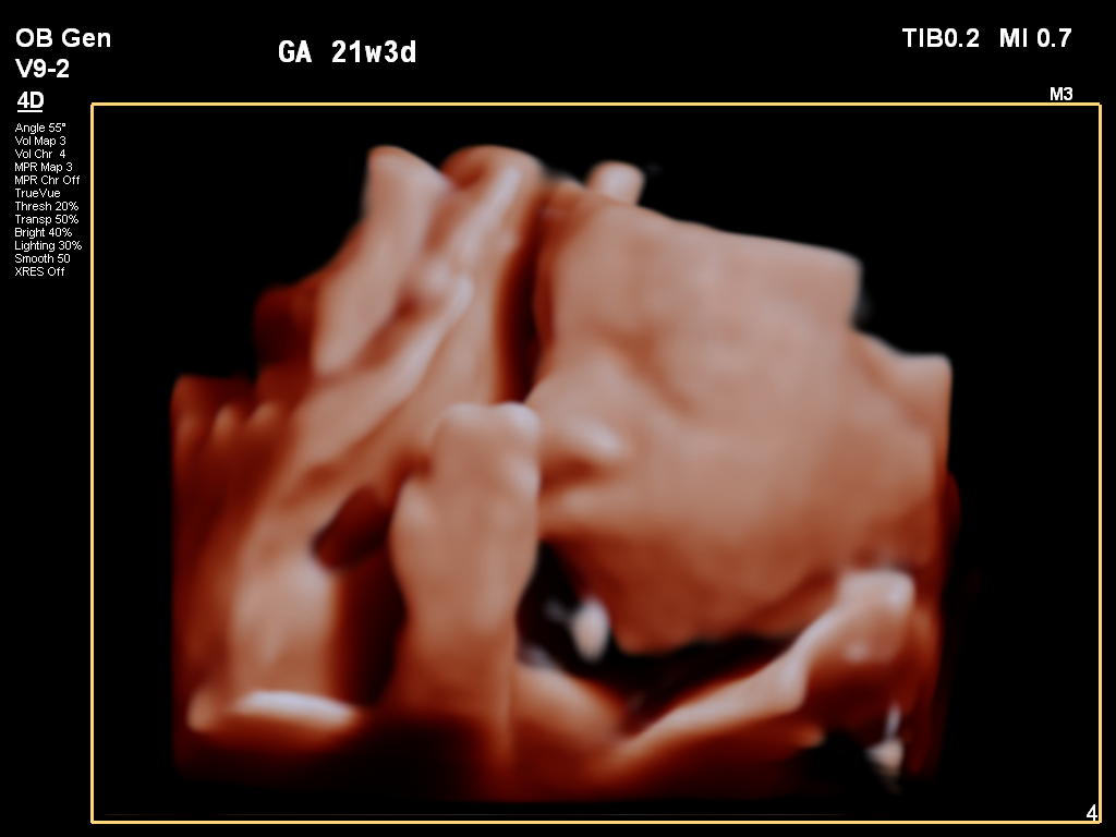
4D ultrasound at 21 weeks 3 days
4D sonography

4D fetal imaging at 21w3d

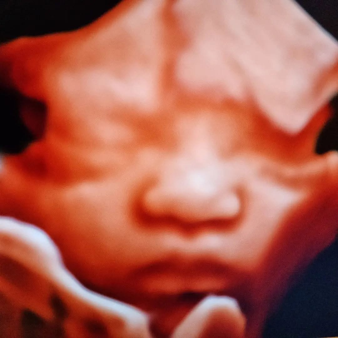
4D ultrasound of a fetal face
4D sonography

Fetal face in 4D

{{ stars }}

“{{ t.quote }}”

{{ t.name }}
{{ t.detail }}
Read & write reviews on Google

Request received

Your request opened in WhatsApp — hit send there and we'll confirm your {{ doneSlot }} slot shortly. If it didn't open, call 89995 54391.

Book another
Rate
{{ selectedRateText }}
Time slot
{{ submitLabel }}
{{ formError }}
Pre-natal sex determination is NOT done here — it is prohibited & punishable by law.

Visit us

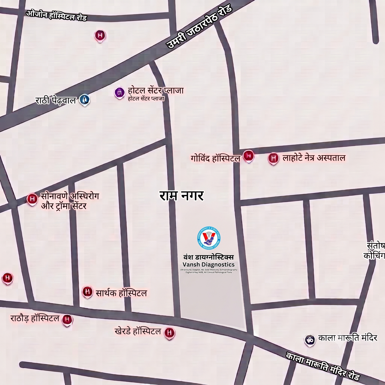
Ground Floor, 166 Main Road, Near Auto Land, Ram Nagar, Akola – 444001
89995 54391 vanshscans@gmail.com
Mon–Sat 9am–8pm · Sun 10am–1pm

Our latest posts load here once the Instagram connection is live. In the meantime, follow us below.

Work with us

We're always glad to hear from sonography technicians, X-ray technicians, receptionists and nursing staff. Send your CV and we'll get in touch when a role opens.

Email your CV

Associate as a doctor

Consultants — radiologists, paediatricians, cardiologists, gynaecologists — interested in visiting or referral association with Vansh Diagnostics are welcome to reach out.

Write to Dr. Sonam